|
MIANYANG XIANFENG MEDECAL INSTRUMENT CO.,LTD
|
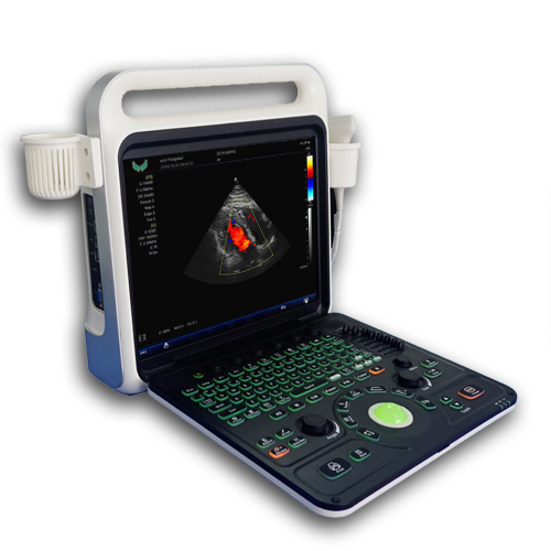


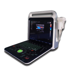
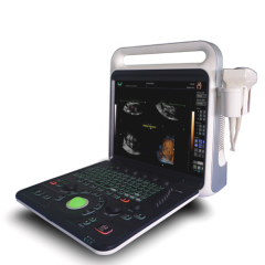
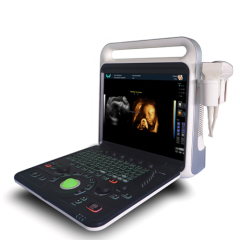
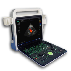
Echowing80 high end color doppler ultrasound diagnostic system
| Price: | 6.0 USD |
| Payment Terms: | T/T,L/C,WU,Paypal,Money Gram |
| Place of Origin: | Sichuan, China (Mainland) |
|
|
|
| Add to My Favorites | |
| HiSupplier Escrow |
Product Detail
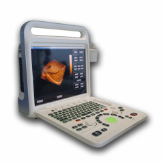
Portable full digital color doppler ultrasound scanner, with high end technical imaging system.Support 3D and 4D imaging.It has high durability
E80 high-end portable color doppler ultrasound diagnostic system,It has unique elegant appearance humanized ergonomic design.This model adopt recent reverse harmonic imaging techlonogy,also able to upgrade the hardware or software(3D or 4D imaging system).The clinical diagnostic performance is better, which can meet the extensive clinical diagnosis needs and create greater value for hospitals.
• 15-inch LCD high resolution displayer
• Reverse tissue harmonic imaging techlonogy
• Multiple channel and beam digital focues techlonogy
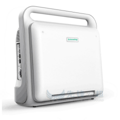
• Compact appearance, easy to carry
• Flexible lifting panel
• Two-level backlit keyboard
• Automitic measurement and analysis vascular intima thickness
• Broadband frequency shift harmonic imaging
It can effectively reduce noise and improve contrast resolution
• Intelligent speckle noise suppression imaging
Intelligent speckle noise suppression technology can intelligently identify different tissue information in different spatial dimensions, inhibit the display of speckle noise edge information, and make the image more exquisite
• Intelligent space composite imaging
Intelligent spatial composite imaging technology, through spatial multi-angle deflection scanning, updates echo signals received from different angles in real time, continuously updates fusion imaging, effectively inhibits random noise and clutter signals under the premise of guaranteeing the temporal resolution, enhances the target organization display, and improves the spatial resolution of the image.High resolution medical liquid crystal display.
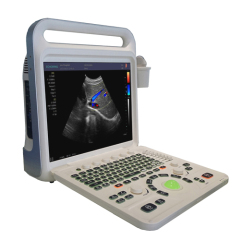
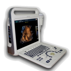
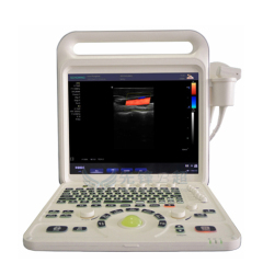
E80 Portable Color Doppler Ultrasound Diagnostic System

Main Functions:
B/2B/4B/M/CFM/PDI/PW;
THI(Tissue Harmonic Imaging);
Real-time 2D and Color Dual Mode;
Real-time 2D and Doppler Double Synchronization;
Real-time 2D ,Doppler and Color triple synchronization;
Free hand 3D software reconstrution imaging(Optional);
Four-dimensional Imaging (4D) (Optional);
High Line Density scan mode for better resolution;
8 sliders TGC Control;
Dynamic range>240 dB;
Overall gain control;
M - mode sweep speed control;
Ultrasound power control;
Variable frame averaging;
Advanced gamma control;
Scan direction, rotation, up-down controls;
Negative / positive control;
Echo enhancement control;
Noise rejection function;
Speckle reduction;
Extended imaging;
Frequency Compound Imaging;
Probe Interface:Two Active Probe Interface;
Multimedia and peripheral devices:Compact Disc-Recordable,USB portable storage device;
Digital Printer / Video Printer / Laser Printer / Ink-Jet Printer;
Memory Function:Image Storage,Video Storage,Cineloop(≥300 Fram),Solid hard disk storage space≥120G;
Puncture guide line:All kinds of probes can register puncture guide line function;
Automatic measurement and analysis of vascular intima;
The puncture guide line Angle adjustable and can be pre-defined;
Language:Chinese, English, Russian, Spanish, Portuguese, French;
Having a full digital beam forming technology
Scanning mode: Convex array, lumen, high-frequency linear array, phased array;4D probe(Optional)
Main Feature:
Systemic applications include abdomen, obstetrics, gynecology,blood vessels, small organs, urology, neonates and pediatrics. Includes scanning modes such as B / CFM / PDI / PW / CW / M and provides excellent resolution and sensitivity. Support convex array probe, linear array probe, convex probe, cavity probe, phased array probe, 4D probe. Integrated network ultrasound workstation, support for DICOM transmission. Using embedded computing system, the system is safe, stable and high-speed operation into one.
Measurement / Calculation
--General Measurement:
--B mode: distance, area (track method, rectangle method, ellipse method), perimeter (track method, rectangle method, ellipse method), volume (double plane method, area length method, ellipsoid method), angle, ratio, stenosis rate.
--M Mode:Slope, ratio, stenosis rate, heart rate, time.
--PW Mode:Doppler blood flow measurement, velocity, acceleration, pressure gradient, time, velocity integral, pulsatility index, resistance index.

--Gynecological measurement and analysis
Uterus, endometrium, ovary, cervix, follicle measurement.
--Obstetric measurement and analysis:
GS gestational sac, CRL head and hip diameter, LV spine length, BPD biparietal diameter, OFD Occipital bone frontal bone diameter, HC head circumference, TAD transverse abdominal diameter, LVW lateral ventricular posterior angle width, HW cerebral hemisphere width, TCD cerebellar lateral diameter , IOD intraocular diameter, OOD external eye distance, BD binocular distance, APTD DBH, TTD transverse DBH, AC abdominal circumference, APD antecedent abdominal diameter, FTA trunk cross-sectional area, HL humerus length, ULNA ulna length, RAD radial length, FL Length of femur, length of tibia, length of fibula of FIB, multiple calculation of APTDxTTD, length of clavicle of clavicle, dysplastic hip (measured hip angle), etc. The gestational age, fetal weight and expected date of birth can be calculated.
--Urology measurement and analysis:
Prostate volume, bladder volume, residual urine volume, volume of prostate transitional area, hip angle measurement and assessment (neonatal hip dislocation diagnosis), cross-sectional measurement (V-Slice method).
--Small Organs and Peripheral Blood Vessel Measurement and Analysis:
The main measurement and analysis of vascular cross-sectional area, heart rate, per volume, flow per unit time, ejection time, stenosis rate, average velocity of blood flow, RI resistance index, PI pulsatile index.
--Measurement report:
Obstetric measurement report, gynecological measurement report, cardiac measurement report, urinalysis and other measurement reports. Automatically store measurement results and generate reports
--symbol:
≥95 kinds of symbol position markers with probe position.
Through the intuitive body mark detail interface quickly select the body mar.
Text markup, presettable text content.
Arrow mark, support multiple arrow marks and adjustable direction.
E80 Portable Color Doppler Ultrasound Diagnostic System

Main Technical Indexes:
The performance requirements of gray-scale imaging mode
The color ultrasonic at the gray-scale imaging performance mode should comply with the provisions of the
table 2.1
Table 2.1 At the Gray-scale imaging mode the performance of the probe
performance indexes | probe type and nominal frequency | |||
2.0≤f<5.0 | 2.0≤f<6.0 | 5.0≤f<10.0 | 5.0≤f<12.0 | |
a) probe type and model | phased array (D4-3D16) | Convex array (C5-2/ R60) | Cavity (EV9-4R10) | Linear array L13-5L53) |
b) nominal frequency (MHz) | 3.0 or 5.0 | 3.5 | 6.5 | 7.5 |
c) Scan depth(mm) | ≥140 | ≥160 | ≥40 | ≥50 |
d) Lateral resolution (mm) | ≤3(depth≤80) ≤4(80<depth≤130) | ≤3(depth≤80) ≤4(80<depth≤130) | ≤2(depth≤30) | ≤2(depth≤40) |
e) Axial resolution (mm) | ≤2(depth≤80) | ≤2(depth≤80) ≤3(80<depth≤130) | ≤1(depth≤40) | ≤1(depth≤50) |
f) Blind area (mm) | ≤7 | ≤5 | ≤4 | ≤3 |
g) Transverse geometry precision (%) | ≤20 | ≤15 | ≤10 | ≤10 |
h) Longitudinal geometric location accuracy (%) | ≤10 | ≤10 | ≤5 | ≤5 |
i) Slice thickness (mm) | ≤5 | ≤5 | ≤5 | ≤5 |
j) Perimeter and area measured deviation (%) | ≤±20 | ≤±20 | ≤±20 | ≤±20 |
k) M mode time display error (%) | ≤±10 | ≤±10 | ≤±10 | ≤±10 |
The performance requirements of color Doppler imaging mode
a). The color ultrasonic at the color Doppler imaging mode should comply with the provisions of the
table 2.2;
b). Color blood flow image should be essentially coincident with the gray-scale image of pipe's;
c). Blood flow direction should be able to correctly identify, no aliasing phenomenon;
Table 2.2 at the color blood flow imaging mode the performance of the probe
Doppler model | Phased array | Convex array | Cavity | Linear array |
Investigation depth at Color blood flow model | ≥90mm | ≥100mm | ≥40mm | ≥50mm |
Investigation depth at Doppler spectrum model | ≥90mm | ≥100mm | ≥40mm | ≥50mm |
Blood flow speed reading error | ≤±15% | |||
The performance requirements of Doppler spectrum mode
a). The color ultrasonic at the color Doppler spectrum mode should comply with the provisions of the;
b). Blood flow speed reading error should comply with the provisions of the table2.3;
c). Pulse wave Doppler mode sampling area cursor position should be accurate;
Table 2.3 at the color blood flow imaging mode the performance of the probe
Doppler model | Phased array | Convex array | Cavity | Linear array |
Investigation depth at Color blood flow model | ≥90mm | ≥100mm | ≥40mm | ≥50mm |
Investigation depth at Doppler spectrum model | ≥90mm | ≥100mm | ≥40mm | ≥50mm |
Blood flow speed reading error | ≤±15% | |||

Displayer: 15 inch LCD color display;
Running hours: ≥8h;
Input power: ≤320V;
Host weight: about 7 kg;
Host appearance size:380 ×212×390(length × width × height) (mm3).
Related Search
Find more related products in following catalogs on Hisupplier.com
Company Info
MIANYANG XIANFENG MEDECAL INSTRUMENT CO.,LTD [China (Mainland)]
Business Type:Manufacturer, Trading Company, Service
City: Mianyang
Province/State: Sichuan
Country/Region: China (Mainland)