|
MIANYANG XIANFENG MEDECAL INSTRUMENT CO.,LTD
|


Full digital color doppler ultrasonic system.3D Real -time imaging
| Price: | 0.1 USD |
| Payment Terms: | T/T,L/C,WU,Paypal,Money Gram |
| Place of Origin: | Sichuan, China (Mainland) |
|
|
|
| Add to My Favorites | |
| HiSupplier Escrow |
Product Detail
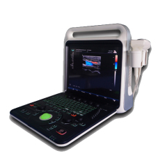
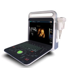
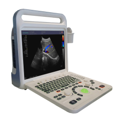
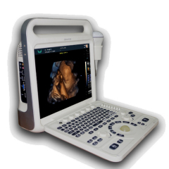
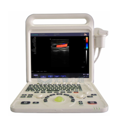
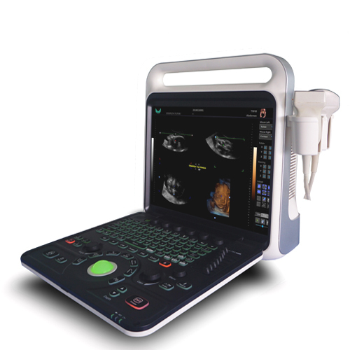
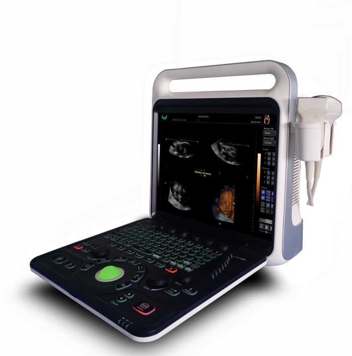
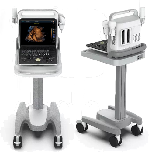
Portable full digital color doppler ultrasound scanner, with solid state drive and stable XP system.Support 3D and 4D imaging.It has high durability.
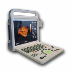
XF3800(4D real-time imaging) color doppler ultrasound system adopts the imaging technology of XF medical high-end products, It has elegant appearance.with excellent image quality, efficient operation flow and high cost performance, which can meet the extensive clinical diagnosis needs and create greater value for hospitals.Humanized ergonomic design.
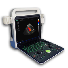
• 15-inch LCD display with up and down adjustment
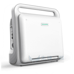
• Compact fuselage, easy to move
• Flexible lifting panel
• Two-level backlit keyboard, more convenient to operatelt can meet the needs of clinical
diagnosis and improve the confidence of diagnosis.
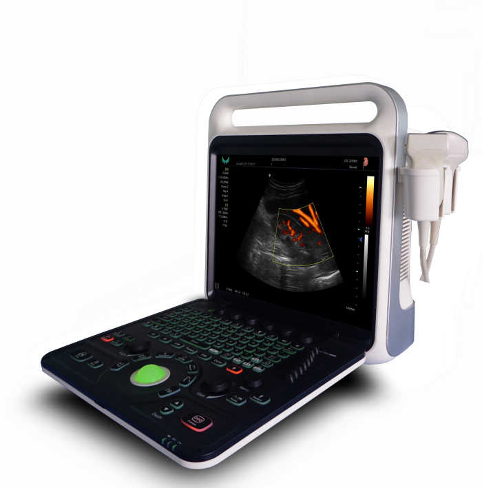
• Broadband frequency shift harmonic imaging
It can effectively reduce noise and improve contrast resolution
•Intelligent speckle noise suppression imaging
Intelligent speckle noise suppression technology can intelligently identify different tissue information in different spatial dimensions, inhibit the display of speckle noise edge information, and make the image more exquisite
•Intelligent space composite imaging
Intelligent spatial composite imaging technology, through spatial multi-angle deflection scanning, updates echo signals received from different angles in real time, continuously updates fusion imaging, effectively inhibits random noise and clutter signals under the premise of guaranteeing the temporal resolution, enhances the target organization display, and improves the spatial resolution of the image.High resolution medical LCD.
Main Functions:
Having a full digital beam forming technology
Scanning mode: Convex array, lumen, high-frequency linear array, phased array;4D probe(Optional)
Probe parameters
*Convex: R60 (R50) Center frequency 3.5MHz (range: 2-6MHz)
*Linear: Center Frequency 7.5MHz (Range: 5-12MHz)
*Cavity probe(TVS): center frequency 6.5MHz (range: 5-10MHz)
*Micro-convex: center frequency 3.5MHz (range: 3-6MHz)
*High-frequency micro-convex probe: the center frequency of 5MHz (range: 4-8MHz)
*Phased array probe center frequency 3Mhz(range:2-5Mhz)
*4D probe : Center frequency 3 MHz (range: 2-5MHz)
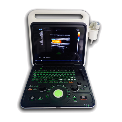
XF3800(4D) Portable full digital color doppler ultrasonic system
--Dynamic range: 0~120dB adjustable;
--Display mode: B,B/B,M,B/M,CFM,CMF/B,PDI,B/PW,CW etc mode;
--Application mode: abdomen, kidneys, urinary system, obstetrics, gynecology, pelvic, small organ, muscle tissue,
organ, breast, heart and other 11 kinds of models;
--Image mode: digital beam forming, tissue harmonic imaging;
--Acoustic output: Mechanical index and thermal index real-time display;
--Acoustic power:Step is adjustable, real-time display;
--Gray scale: 256 scales;
--Depth display: ≥250mm;
--B/D triple-purpose: linear array: B/PW D; convex array: B/PW D;
--Pseudo color processing: 16 kinds of pseudo color encoding can optional;
--Gain adjusts: 8 segments TGC, B/M/D/C is independently adjustable; TGC curve can show and hide automatically;
--Image magnification: picture in picture zoom in and zoom part function;
--Self-motion optimize function: Built-in multiple check type, according to different inspection organs, preset best image check condition, reduce the adjusting operation keys;
--One-click optimization function: preset several parameters adjusting focus on a button, a key to realize image
fast optimization;
Measurement and calculation:
--Distance, circumference, area, volume, angle, ratio, and stenos rate.
--M mode routine measurement: Heart rate, time, distance, speed, ratio, etc.
--Gynecology measurement: Uterus, cervix, endometrial, ovary, follicular.
Obstetrics measurement:
EGA, ETD, fetal weight estimation, AFI index, OB report (including OB tables).
Cardiology measurement: LV measurement.
Urology measurement:
Prostate volume, displacement volume, bladder capacity, and residual urine output.
PW measurements: Time, speed, Heart Rate, RI, PI, etc.
Other measurement:
Slice volume measurement, hip joint angle measurement.
Image storage: Image storage, video storage, cine loop, disk storage capacity≥160G;
Patient data: Medical record management, report inquiry and printing, image video output( HDD ,USB,Optional
DVD-RW),built-in ultrasound workstation;
Reporting system:
automatic report generation system, and can be full screen characters in both Chinese and
English editor;
Output interface:SR323,USB,DICOM interface;
XF3800(4D) Portable full digital color doppler ultrasonic system
Main Technical Indexes:
The performance requirements of gray-scale imaging mode
The color ultrasonic at the gray-scale imaging performance mode should comply with the provisions of the
table 2.1
Table 2.1 At the Gray-scale imaging mode the performance of the probe
performance indexes | probe type and nominal frequency | |||
2.0≤f<4.0 | 2.0≤f<5.0 | 5.0≤f<8.0 | 5.0≤f<10.0 | |
a) probe type and model | phased array (type TP16A) | Convex array (type TC60A) | Cavity (type TC10A) | Linear array (type TL40A) |
b) nominal frequency (MHz) | 3.0 | 3.5 | 6.5 | 7.5 |
c) Scan depth(mm) | ≥140 | ≥160 | ≥40 | ≥50 |
d) Lateral resolution (mm) | ≤3(depth≤80) ≤4(80<depth≤130) | ≤3(depth≤80) ≤4(80<depth≤130) | ≤2(depth≤30) | ≤2(depth≤40) |
e) Axial resolution (mm) | ≤2(depth≤80) | ≤2(depth≤80) ≤3(80<depth≤130) | ≤1(depth≤40) | ≤1(depth≤50) |
f) Blind area (mm) | ≤7 | ≤5 | ≤4 | ≤3 |
g) Transverse geometry precision (%) | ≤20 | ≤15 | ≤10 | ≤10 |
h) Longitudinal geometric location accuracy (%) | ≤10 | ≤10 | ≤5 | ≤5 |
i) Slice thickness (mm) | ≤5 | ≤5 | ≤5 | ≤5 |
j) Perimeter and area measured deviation (%) | ≤±20 | ≤±20 | ≤±20 | ≤±20 |
k) M mode time display error (%) | ≤±10 | ≤±10 | ≤±10 | ≤±10 |
The performance requirements of color Doppler imaging mode
a). The color ultrasonic at the color Doppler imaging mode should comply with the provisions of the
table 2.2;
b). Color blood flow image should be essentially coincident with the gray-scale image of pipe's;
c). Blood flow direction should be able to correctly identify, no aliasing phenomenon;
Table 2.2 at the color blood flow imaging mode the performance of the probe
Doppler model | Phased array | Convex array | Cavity | Linear array |
Investigation depth at Color blood flow model | ≥90mm | ≥100mm | ≥40mm | ≥50mm |
Investigation depth at Doppler spectrum model | ≥90mm | ≥100mm | ≥40mm | ≥50mm |
Blood flow speed reading error | ≤±15% | |||
The performance requirements of Doppler spectrum mode
a). The color ultrasonic at the color Doppler spectrum mode should comply with the provisions of the;
b). Blood flow speed reading error should comply with the provisions of the table2.3;
c). Pulse wave Doppler mode sampling area cursor position should be accurate;
Table 2.3 at the color blood flow imaging mode the performance of the probe
Doppler model | Phased array | Convex array | Cavity | Linear array |
Investigation depth at Color blood flow model | ≥90mm | ≥100mm | ≥40mm | ≥50mm |
Investigation depth at Doppler spectrum model | ≥90mm | ≥100mm | ≥40mm | ≥50mm |
Blood flow speed reading error | ≤±15% | |||
Displayer: 15 inch LCD color display;
Running hours: ≥8h;
Input power: ≤320V;
Host weight: about 7 kg;
Host appearance size:380 ×212×390(length × width × height) (mm3).
Related Search
Find more related products in following catalogs on Hisupplier.com
Company Info
MIANYANG XIANFENG MEDECAL INSTRUMENT CO.,LTD [China (Mainland)]
Business Type:Manufacturer, Trading Company, Service
City: Mianyang
Province/State: Sichuan
Country/Region: China (Mainland)